Abstract
Introduction: Ultrasound and CT angiography are common diagnostic methods of abdominal aortic pathologies. In the last decade, hybrid methods (PET/CT, PET/MRI) have become more common in this diagnostic algorithm. Originally they were indicated in malignancies or inflammatory processes. Currently, efforts are developed to visualize possible local inflammatory activity in the aortic wall and thus to assess a certain “disease activity” with the goal to anticipate further development of aortic pathology. The aim of our study was to analyze potential benefits of hybrid methods in predicting abdominal aortic pathology progression.
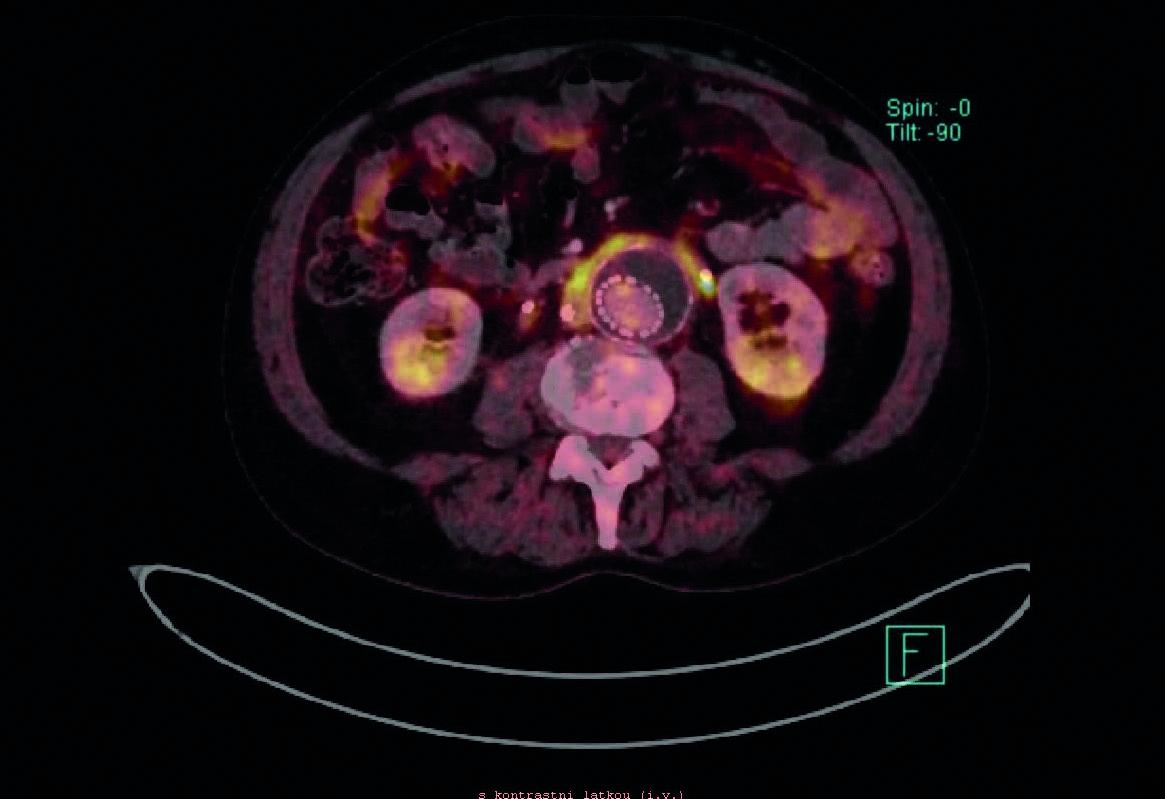
Methods: In this prospective, open-label, observational study we examined 75 patients referred to PET/CT (N=61) or PET/MRI (N=14) due to any aortic pathology in 2015–2017. The patients included those with abdominal aortic aneurysm (AAA) (N=48; 64%), aortitis (N=5; 6.7%), aortic dissection (N=4; 5.3%), patients undergoing EVAR (N=6; 8%), patients with excessive atherosclerosis (N=7; 9.3%), patients with concomitant AAA and retroperitoneal fibrosis (N=4; 5.3%) and patient with an intramural hematoma (N=1; 1.3%). The minimum follow-up period was 6 months (0.5–2.5 years). Clinical symptoms, aortic diameter, growth rate and CRP levels were analyzed during the follow-up and correlation with PET/CT or PET/MRI findings was evaluated.
Results: Increased metabolic activity in the aorta was found in 25 of the 75 examined patients (33.3%). Based on statistical analysis there were no associations between increased activity based on PET/CT or PET/MRI in the aortic wall and disease symptoms or progression.
Conclusion: Our results provide no evidence that hybrid methods can predict further development of pathological findings...

