Abstract
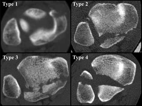
The study presents an overview of the most common radiography and CT-based classifications of posterior malleolar fractures in ankle fracturedislocations. Their analysis has shown that posterior malleolar fractures largely vary in size and shape. Evaluation of fractures by plain radiographs is inadequate. A detailed assessment of the fragment shape and course of fracture lines requires CT examination in all three projections, followed by 3D CT reconstructions.

