Abstract
Introduction: Early diagnosis of complicated healing of colorectal anastomosis can increase the chance for salvage surgery and thus reduce overall morbidity. Confocal laser endomicroscopy (CLE) enables in vivo assessment of tissue perfusion without disturbing its integrity. This experimental study evaluates the potential of CLE for postoperative monitoring of colorectal anastomosis.
Methods: A hand-sewn colorectal anastomosis was performed in 9 pigs. The animals were subsequently divided into groups with normal (N=3) and ischemic anastomosis (N=6). Microscopic signs of hypoperfusion were evaluated postoperatively at regular intervals using CLE.
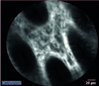
Results: Uneven saturation of the images was evident in the group with ischemic anastomosis. The epithelium had inhomogeneous edges and more numerous crypt branching was visible. Tissue oedema quantified as the number of crypts per visual field was already more extensive at the first measurement after induction of ischemia. There was also a significant difference between the values measured before and 10 minutes after ischemia – 8.7±1.9 vs. 6.0±1.1 (p=0.013).
Conclusion: Postoperative monitoring of the colorectal anastomosis using CLE enables prompt detection of perfusion disorders

