
Keywords
rectosigmoid adenocarcinoma
renal-cell carcinoma
prostate adenocarcinoma
neuroendocrine small-cell lung cancer
Abstract
Introduction: Multiple tumors belong to rare cancers. Almost all malignancies may occur in combination, the most common combination being gastrointestinal cancer with respiratory or urogenital tract cancer and with breast cancer in females.
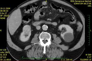
Case report: In 1999, a 66-year-old patient was diagnosed with a rectosigmoid tumor histologically proven as adenocarcinoma. Rectosigmoid resection was performed, followed by adjuvant radiotherapy and the Mayo Clinic FU/FA chemotherapy regimen. Radical nephrectomy was performed in January 2005 due to right kidney tumor, histologically detected as clear cell carcinoma. In February 2006, the patient underwent bilateral pelvic lymphadenectomy for biopsy-verified adenocarcinoma of the prostate with PSA 20.8 ng/ml. Radical prostatectomy was performed in April 2006. Histology demonstrated moderately differentiated adenocarcinoma in both prostatic lobes classified as Gleason score 4 (1+3), without invasion into the capsule or seminal vesicles infiltration. In June 2016, a native X-ray of the lungs revealed a subpleural small dense node in the right upper pulmonary field. PET/CT of the trunk was also performed showing liver metastasis and pulmonary deposits, including enlargement of the mediastinal nodules. In October 2016, liver biopsy was taken and the serum level of neuron-specific enolase (NSE: 93 ng/ml) was measured. Histology demonstrated neuroendocrine carcinoma of the small cell type. In November 2016, palliative chemotherapy with carboplatin and etoposide administered once a month was initiated. After 4 chemotherapy cycles, no deposits on the liver were detected by sonography. A native X-ray image of the lungs still showed a 15mm deposit, but NSE levels returned to normal. In March 2017, treatment continued with palliative radiotherapy of the right lung, mediastinal lymphatic nodes and prophylactic radiotherapy of the skull was planned as a next step. In August 2017, the patient died due to renal function failure and deterioration of the general condition.
Conclusion: The patient worked in uranium mines and underwent radio-chemotherapy after the first malignancy – rectosigmoid tumor. Genetic examination was not performed. The patient died of therapeutic complications of the last malignancy. Our case report does not confirm findings described so far – a shortening interval between malignancies – but confirms their increasing aggressiveness.