Abstract
Introduction: Multidisciplinary management of patients with rectal cancer presents a gold standard of care; neoadjuvant therapy indications are based on magnetic resonance imaging (MRI) description of the local stage of the carcinoma. Although the accuracy of MRI-based assessment of cancer depth of invasion is satisfactory, its accuracy in the assessment of mesorectal lymphadenopathy is very questionable.
Methods: This was a prospective, single-centre, cohort study focused on the accuracy of preoperative MRI in the assessment of mesorectal lymph nodes (LN). MRI findings of each patient were compared with detailed histopathological examination of rectal specimens.
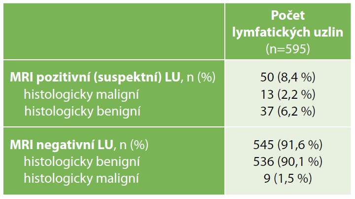
Results: Forty patients with rectal cancer, undergoing rectal resection with total mesorectal excision were enrolled in the study. MRI assessment of the T-stage was correct in 22 of the 40 study patients (55.0%). T-stage overstaging was noted in 14 (35.0%), and understaging in 4 (10.0%) study patients. According to preoperative MRI (using Horvat’s criteria), there were 50 suspicious/malignant lymph nodes. Only 13 of these 50 LNs (26.0%) were proved malignant on histopathology examination. In total, our study group included 18 patients with suspicious/positive LNs (according to preoperative MRI) who were classified as cN+. MRI diagnosis of malignant lymphadenopathy was correct in only 33.3% of these patients.
Conclusion: MRI shows very low accuracy in the evaluation of mesorectal lymph nodes in patients with rectal cancer. Therefore neoadjuvant therapy should be offered particularly with respect to MRI description of the depth of carcinoma invasion (T-stage and relationship to fascia propria of the rectum).

