
Klíčová slova
Abstrakt
Introduction: Na experimentálnu verifikáciu vitality žalúdočného tubulu pred ezofagektómiou je nevyhnutná existencia jednoduchého modelu. Ischémia je hlavná príčina dehiscencie ezofagogastrickej anastomózy a následného leaku anastomózy. Ischemická príprava žalúdočného tubulu pred ezofagektómiou je potencionálna možnosť ako zamedziť rozvoju nežiaducich komplikácií. Avšak technika ako aj optimálne načasovanie ischemizácie ostávajú nejasné.
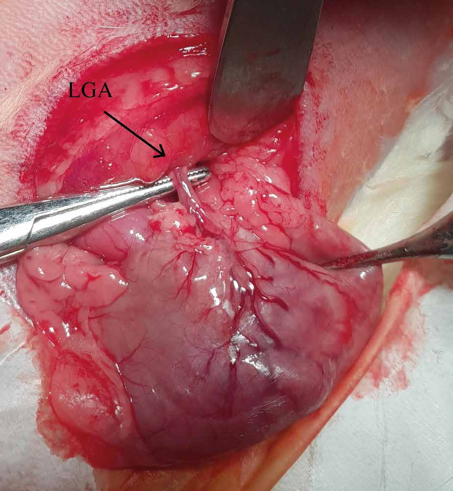
Metody: Samce potkanov plemena Sprague-Dawley (n=24) boli náhodne rozdelené do štyroch skupín: ischemická skupina s odberom vzoriek po 1 hodine od ischémie (I1H), ischemická skupina s odberom vzoriek 1 deň od ischémie (I1D), ischemická skupina s odberom vzoriek 7 dní od ischémie (I7D) a kontrolná skupina (C). Ischémia bola navodená ligáciou a. gastrica sinistra a aa. gastricae breves. Vzorky boli histologicky a makroskopicky verifikované a stanovený bol počet a percentuálne zastúpenie jednotlivých imunokompetentných buniek.
Výsledky: V skupine I1H bola pozorovaná ischemická denudácia s mukozálnymi eróziami a celkový počet eozinofilov bol signifikantne vyšší (p<0.05) v tejto skupine v porovnaní so skupinami I1D a I7D. V skupine I1D bola pozorovaná redukcia zápalu a parciálna regenerácia žalúdočnej mukózy. V skupine I7D bola pozorovaná takmer kompletná architektonická regenerácia žalúdočného epitelu a počet polymorfonukleárov bol signifikantne nižší (p<0,05) ako v skupine I1D.
Záver: Ischemické poškodenie ligáciou žalúdočných tepien bolo predominantne pozorované v skupinách I1H a I1D avšak nie v skupine I7D. Táto práca prezentuje jednoduchú metódu verifikácie ischemických zmien žalúdočného tubulu.