
Keywords
extramural vascular invasion
progression-free survival
Abstract
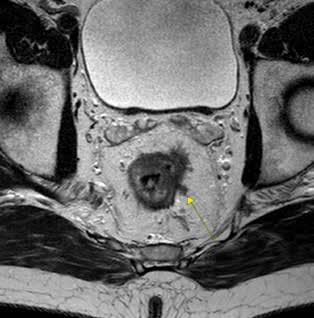
Introduction: Extramural vascular invasion (EMVI) is associated with a poor prognosis in patients with rectal carcinoma. Patients with proven vascular invasion have a shorter progression-free survival and overall survival. Until recently, vascular invasion had been identified primarily by pathologists. Currently, EMVI can be detected preoperatively by magnetic resonance imaging used for rectal cancer staging. Our study aimed at verifying the effect of pre-operative EMVI detection on PFS after resection and comparing this interval (PFS) to the group of patients with vascular invasion identified and confirmed by pathologists.
Methods: Patients who underwent surgery for rectal carcinoma at our Surgical Department in the years 2012−2016 were included in the group and were followed for local recurrence or systemic progression of the disease. The median follow-up was 36 months. In this group, we then retrospectively evaluated MR EMVI and at the same time the presence of tumor vascular invasion from the resected specimen. The relationship of both prognostic markers to PFS was compared.
Results: Tumor vascular invasion as well as positive extramural vascular invasion on MRI found preoperatively in our group had a statistically significant negative effect on the progression-free survival compared to the group without evidence of EMVI or vascular invasion.
Conclusion: Positive extramural vascular invasion found on MRI during rectal cancer staging is associated with a poor prognosis. It is one of the prognostically negative factors and referral of these patients for outpatient care should receive special attention because even after radical resection with a negative resection line there is a risk of early progression of the disease.